AUTORES
Vitório Campos da Silva
Cirurgião-dentista – Faculdade de Odontologia João Prudente/FAEE; Doutor em Patologia Celular e Molecular, e pós-doutorando em Medicina – Universidade de Brasília.
Orcid: 0000-0002-7968-6951.
Gustavo Henrique Takano Soares
Médico patologista docente – Faculdade de Medicina da Universidade de Brasília.
Orcid: 0000-0001-5553-3582.
Brenda de Pina Campos de Medeiros
Cirurgiã-dentista e especialista em Ortodontia – Ipesp, Brasília; 1ª Ten. QOCON-HMAB – Exército Brasília/DF.
Bruna Campos de Freitas
Cirurgiã-dentista e especialista em Radiologia – ABO/DF.
RESUMO
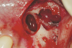
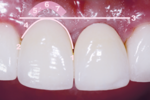
A procura por biomateriais substitutos ósseos começou há muito tempo. Embora o osso autógeno medular seja considerado o padrão-ouro e o tecido ósseo possua a capacidade de se autorregenerar, há situações em que o uso dos biomateriais sintéticos é imprescindível. O objetivo deste trabalho foi utilizar a microscopia eletrônica de varredura (MEV) na avaliação topográfica das superfícies do Nanosynt, além de observar in vitro a interação de granulócitos humanos, separados pela técnica do Percoll e cultivados nos grânulos deste biomaterial. Os resultados mostram que tais células interagem com o Nanosynt, sendo ativadas e exibindo alterações morfológicas indicativas da sua adesão à superfície deste material aloplástico. Imagens obtidas com a utilização da MEV duas horas após o cultivo mostram células ativadas na superfície dos grânulos, e imagens da microscopia ótica (MO) mostram ainda células diferenciadas, multinucleadas, reabsorvendo o biomaterial na periferia e nos poros dos grânulos do Nanosynt, três meses após o seu enxerto em maxila humana.
Palavras-chave – Microscopia eletrônica de varredura; Microscopia ótica; Interação celular; Neutrófilos; Nanosynt.
ABSTRACT
There has long been a demand for bone substitute biomaterials. Although autologous bone marrow is considered the “gold standard” and the bone tissue has the capacity to self-regenerate, there are situations where the use of synthetic biomaterials is essential. The objective of this work was to use scanning electron microscopy (SEM) in the topographic evaluation of Nanosynt surfaces and to observe in vitro the interaction of human granulocytes, separated by the Percoll technique and cultured in the granules of this biomaterial. The results show that such cells interact with Nanosynt being activated, exhibiting morphological changes indicative of the adhesion on the surface of this alloplastic material. Two hours after the culturing of the cells, activated cells on the surface of the granules and images of the optical microscopy (OM) show that multinucleated cells reabsorbing the biomaterial, at the periphery and in the pores of Nanosynt granules, three months after its graft in the human jaw.
Key words – Scanning electron microscopy; Optical microscopy; Cell interaction; Neutrophils; Nanosynt.
Recebido em jun/2019
Aprovado em jun/2019